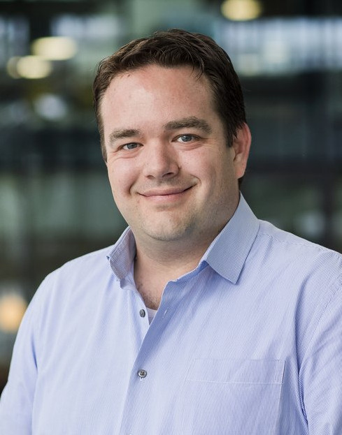
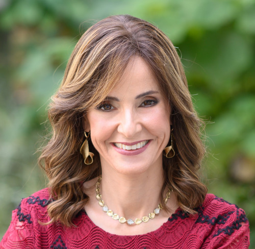
Yonina Eldar
Title: Model based deep learning with applications to inverse ultrasound
Abstract: Deep neural networks provide unprecedented performance gains in many real-world problems in signal and image processing. Despite these gains, the future development and practical deployment of deep networks are hindered by their black-box nature, i.e., a lack of interpretability and the need for very large training sets. On the other hand, signal and image processing have traditionally relied on classical statistical modeling techniques that utilize mathematical formulations representing the underlying physics, prior information and additional domain knowledge. Simple classical models are useful but sensitive to inaccuracies and may lead to poor performance when real systems display complex or dynamic behaviour. Here we introduce various approaches to model based learning which merge parametric models with optimization tools and classical algorithms leading to efficient, interpretable networks from reasonably sized training sets. We will then show how these methods can be used to address quantitative ultrasound by treating channel data as an inverse ultrasound problem.
Bio: Yonina Eldar is a Professor in the Department of Mathematics and Computer Science, Weizmann Institute of Science, Rehovot, Israel, where she heads the center for Biomedical Engineering and Signal Processing and holds the Dorothy and Patrick Gorman Professorial Chair. She is also a Visiting Professor at MIT, a Visiting Scientist at the Broad Institute, a Visiting Research Collaborator at Princeton, an Adjunct Professor at Duke University, an Advisory Professor of Fudan University, a Distinguished Visiting Professor of Tsinghua University, and was a Visiting Professor at Stanford. She is a member of the Israel Academy of Sciences and Humanities and of the Academia Europaea, an IEEE Fellow a EURASIP Fellow, a Fellow of the Asia-Pacific Artificial Intelligence Association, and a Fellow of the 8400 Health Network. She received the B.Sc. degree in physics and the B.Sc. degree in electrical engineering from Tel-Aviv University, and the Ph.D. degree in electrical engineering and computer science from MIT, in 2002. She has received many awards for excellence in research and teaching, including the IEEE Signal Processing Society Technical Achievement Award (2013), the IEEE/AESS Fred Nathanson Memorial Radar Award (2014) and the IEEE Kiyo Tomiyasu Award (2016). She was a Horev Fellow of the Leaders in Science and Technology program at the Technion and an Alon Fellow. She received the Michael Bruno Memorial Award from the Rothschild Foundation, the Weizmann Prize for Exact Sciences, the Wolf Foundation Krill Prize for Excellence in Scientific Research, the Henry Taub Prize for Excellence in Research (twice), the Hershel Rich Innovation Award (three times), and the Award for Women with Distinguished Contributions. She received several best paper awards and best demo awards together with her research students and colleagues, was selected as one of the 50 most influential women in Israel, and was a member of the Israel Committee for Higher Education. She is the Editor in Chief of Foundations and Trends in Signal Processing, a member of several IEEE Technical Committees and Award Committees, and heads the Committee for Promoting Gender Fairness in Higher Education Institutions in Israel.
Note: The talk will be given remotely
Ben Cox

Title: Biomedical Ultrasound Tomography, Choices and Opportunities
Abstract: Ultrasound has been used for biomedical imaging for over 50 years. Recently there has been a revival of interest in tomographic and quantitative methods of imaging with ultrasound. In this talk I will give an overview of biomedical ultrasound imaging methods, highlighting the choices that are made to make them feasible, and pointing to the many opportunities for exciting developments
Bio: Ben Cox is Professor of Biomedical Acoustics at University College London. He is a co-author of the biomedical acoustic modelling toolbox k-Wave, widely used in both academia and industry. He has published over 100 journal articles covering many aspects of photoacoustic tomography and biomedical ultrasound, including numerical and mathematical modelling, instrumentation development, image reconstruction, and other inverse problems.
Christian Böhm

Title: (Full)-Waveform Inversion across the Scales
Abstract: Despite the vastly different scales, ultrasound computed tomography and seismic imaging share remarkable similarities. The increase and availability of computational capabilities in recent years have led both fields to converge and initiated interdisciplinary research. This talk will give an overview of the history, recent advances, and current trends in waveform inversion; highlighting examples for the quantitative imaging of soft tissues and bone using in-vivo data, and how they relate to challenges in seismic tomography.
Bio: Christian Boehm is a Lecturer at the Department of Earth Sciences at ETH Zurich. He obtained his PhD in Mathematics from the Technical University of Munich in 2015. His research focuses on computational inverse problems and wave-based imaging, and he has co-authored over 50 publications with applications in medical ultrasound computed tomography, seismic imaging, and non-destructive testing. He is a chair of the SPIE Medical Imaging Conference on Ultrasonic Imaging and Tomography, and a co-founder of the ETH spin-off Mondaic.
Richard Lopata

Title: High-Fidelity Sonography using Multi-Aperture Imaging
Abstract: Ultrasound imaging is renowned for its high spatio-temporal resolution, low cost, and ease-of-use, but is notorious for its anisotropic image quality and limited field of view. Multi-aperture, multi-static imaging has the potential to overcome several well-known limitations, adapting concepts of US tomography, but for a wide range of applications. In this talk, an overview of our ongoing research in this field will be given, for several applications, from bench to bedside.
Bio: Richard G. P. Lopata received the M.Sc. degree in biomedical engineering from the Eindhoven University of Technology (TU/e), Eindhoven, The Netherlands, in 2004, and the Ph.D. degree from Radboudumc, Nijmegen, The Netherlands, in 2010, with a focus on 2-D and 3-D ultrasound strain imaging: methods and in vivo applications. He has been an Associate Professor at TU/e, heading the Photoacoustics and Ultrasound Laboratory Eindhoven (PULS/e lab) since 2014. The PULS/e lab facilitates research on technology development in the areas of ultrasound functional imaging, photoacoustics, and image-based modeling aimed to facilitate in and/or improve clinical decision-making for cardiovascular, musculoskeletal, and abdominal applications. In recent years he made it his mission to move away from conventional ultrasound using a single handheld transducer to more sophisticated and complex imaging geometries that allow for semi-tomographic, high quality imaging of e.g. the abdomen.


